The Neurotransmitter & Receptor Atlas: A Visual Reference
Every major neurotransmitter and receptor family — structures, synaptic signaling, and clinical drug targets — in one visual reference
Clinical Summary
This atlas is the hub of the PsychoPharmRef neuroscience section, mapping neurotransmitters, their receptor families and localization, the signal-transduction logic that predicts drug effects, and the xanomeline muscarinic mechanism.
Psychopharmacology becomes intuitive the moment you stop memorizing drug lists and start thinking in systems: which neurotransmitter, which receptor, and which signal-transduction pathway. This atlas gathers all of that on one page — the molecules, the synapse, the receptor families, and the exact points where drugs intervene. It is the hub of the neuroscience section; each system below links to a dedicated deep dive.
1 · The Master Map: Neurotransmitters at a Glance
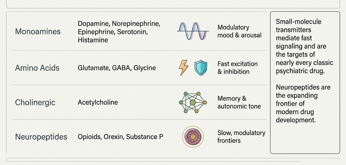
Neurotransmitters fall into a handful of chemical classes. The small-molecule transmitters — monoamines, amino acids, and acetylcholine — mediate fast, moment-to-moment signaling and are the targets of nearly every classic psychiatric drug. Neuropeptides and gasotransmitters act as slower, modulatory co-transmitters and are an expanding frontier of drug development (orexin antagonists for insomnia, opioid modulators for pain and depression).
| Class | Neurotransmitter | Receptor families | Primary functions & clinical relevance |
|---|---|---|---|
| Monoamines | Dopamine (DA) | D₁–D₅ | Reward, motivation, motor control, prolactin. Psychosis, ADHD, addiction, Parkinson’s. |
| Norepinephrine (NE) | α₁, α₂, β₁–β₃ | Arousal, attention, stress response, mood. Depression, anxiety, PTSD, ADHD. | |
| Epinephrine | α, β adrenergic | Peripheral sympathetic (“fight-or-flight”); minor CNS role. | |
| Serotonin (5-HT) | 5-HT₁–5-HT₇ (7 families) | Mood, anxiety, sleep, appetite, GI motility, emesis. SSRIs/SNRIs, antipsychotic 5-HT₂₂₀ effects. | |
| Histamine (HA) | H₁–H₄ | Wakefulness, appetite. H₁ antagonism → sedation and weight gain. | |
| Amino acids | Glutamate | NMDA, AMPA, kainate, mGlu₁–₈ | Main excitatory transmitter; learning & LTP. Ketamine (NMDA), TRD, dementia, excitotoxicity. |
| GABA | GABA₀ (ion), GABAₕ (GPCR) | Main inhibitory transmitter. Benzodiazepines, alcohol, anesthetics, anticonvulsants. | |
| Glycine | GlyR; NMDA co-agonist | Inhibitory in spinal cord/brainstem; obligate NMDA co-agonist. | |
| Cholinergic | Acetylcholine (ACh) | Muscarinic M₁–M₅; nicotinic | Memory, arousal, autonomic tone. Anticholinergic delirium; cholinesterase inhibitors in dementia. |
| Neuropeptides | Opioid peptides | μ (MOR), δ (DOR), κ (KOR) | Analgesia, reward, dysphoria (κ). Opioid use disorder, novel antidepressants. |
| Orexin (hypocretin) | OX₁, OX₂ | Sleep–wake stability. Dual orexin antagonists for insomnia; narcolepsy. | |
| Substance P | Neurokinin NK₁ | Pain transmission, emesis, stress. NK₁ antagonists (antiemetic). | |
| Gasotransmitter | Nitric oxide (NO) | Soluble guanylate cyclase | Retrograde signaling, vasodilation, synaptic plasticity. |
2 · What the Molecules Look Like
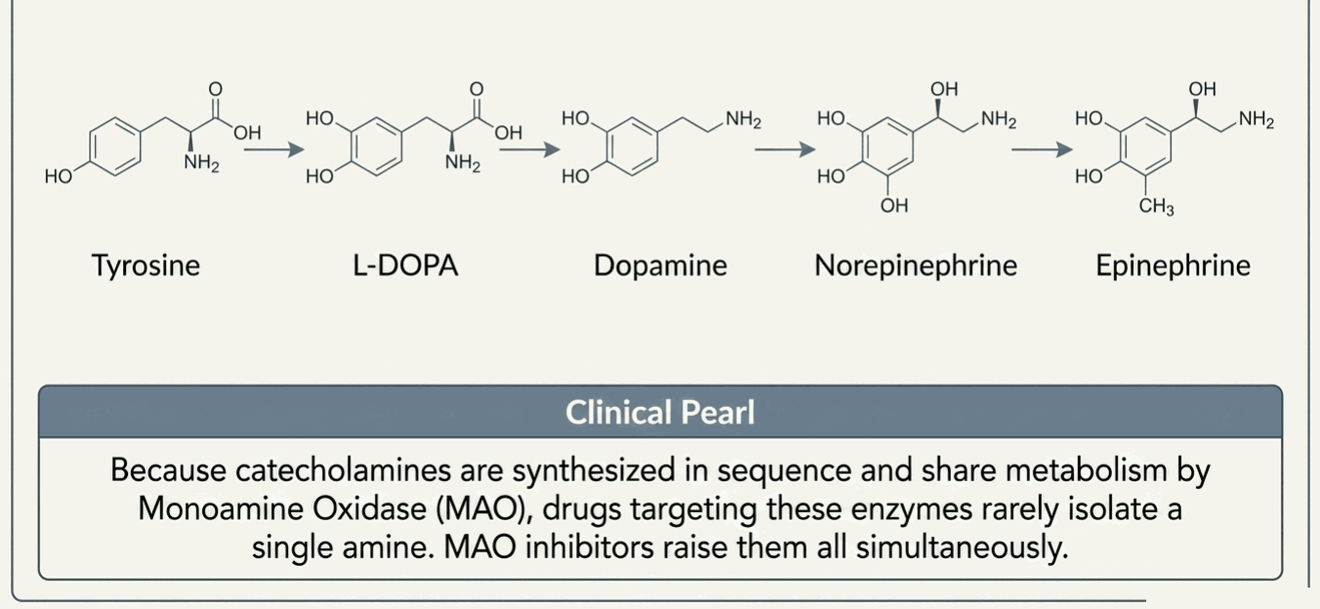
Structure explains kinship. The three catecholamines — dopamine, norepinephrine, and epinephrine — share a catechol ring (benzene with two hydroxyls) and are built sequentially along one synthetic pathway: tyrosine → L-DOPA → dopamine → norepinephrine → epinephrine. Serotonin is an indoleamine derived from tryptophan; histamine derives from histidine. All four monoamines are substrates for monoamine oxidase (MAO), which is why MAO inhibitors raise all of them at once. GABA and glutamate, by contrast, are simple amino acids — glutamate is decarboxylated to GABA by GAD, making them a metabolic parent-and-child pair.
3 · How a Synapse Works — and Where Drugs Act
Every aminergic synapse runs the same production line. A precursor amino acid is converted by synthetic enzymes into the transmitter, which VMAT packages into vesicles. An arriving action potential opens voltage-gated calcium channels (VGCC); calcium triggers vesicle fusion and release into the cleft. Released transmitter then does three things: it activates postsynaptic receptors, it feeds back onto presynaptic autoreceptors to dampen further release, and it is cleared — mostly by a reuptake transporter (SERT, DAT, or NET), with intracellular MAO degrading what returns.
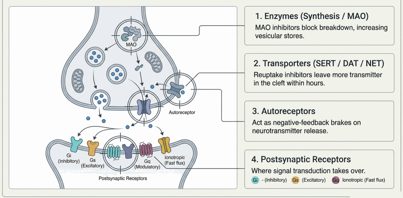
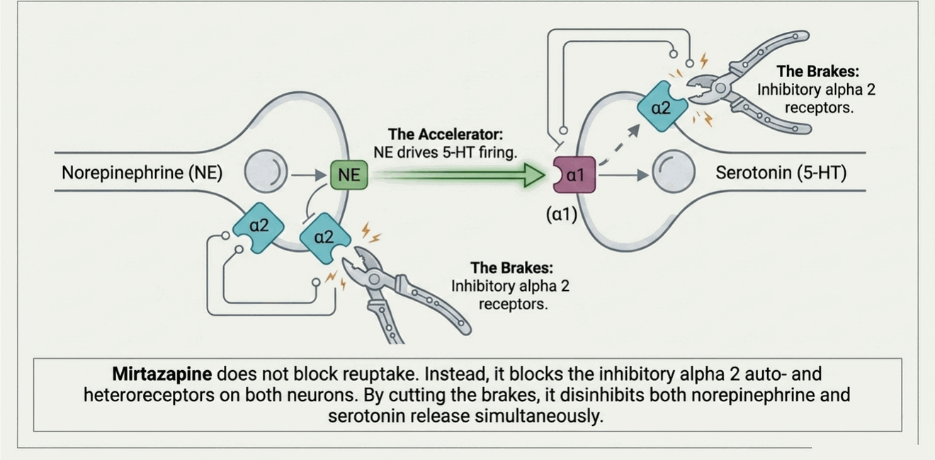
This single picture organizes most of the formulary. Reuptake inhibitors (SSRIs, SNRIs, TCAs, and stimulants at DAT/NET) block the transporter, leaving more transmitter in the cleft. MAO inhibitors block intracellular breakdown, raising vesicular stores. Autoreceptor pharmacology explains delayed and paradoxical effects — mirtazapine’s α₂ blockade disinhibits NE and 5-HT release, and the initial 5-HT₁₀ autoreceptor “brake” is one reason SSRIs take weeks to work. Everything else happens at the postsynaptic receptor, which is where signal transduction takes over.
4 · The Four Signaling Buckets
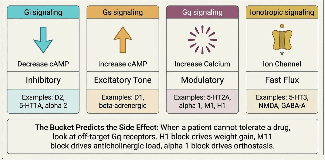
Receptors look bewildering until you realize almost all of them signal through just four mechanisms. Three are G-protein-coupled receptors (GPCRs, or metabotropic — slow, modulatory) and one is the ligand-gated ion channel (ionotropic — fast). Knowing a receptor’s bucket predicts what activating or blocking it will do.
| Receptor | Type / coupling | Signal transduction | Clinical implication (example drugs) |
|---|---|---|---|
| D₂ | GPCR, Gᵢ | ↓ cAMP | Antipsychotic efficacy & EPS; blockade raises prolactin (antipsychotics). |
| D₁ | GPCR, Gₛ | ↑ cAMP | Prefrontal cognition, motor reward; target of interest in cognition. |
| 5-HT₁₀ | GPCR, Gᵢ | ↓ cAMP | Anxiolytic/antidepressant (buspirone partial agonist; vilazodone, vortioxetine). |
| 5-HT₂₀ | GPCR, Gₜ | ↑ IP₃/DAG/Ca²⁺ | Antagonism → atypical antipsychotic action; agonism → psychedelics. |
| 5-HT₂₊ | GPCR, Gₜ | ↑ IP₃/DAG/Ca²⁺ | Antagonism → appetite & weight gain (mirtazapine, olanzapine). |
| 5-HT₃ | Ion channel | Na⁺/Ca²⁺ influx | Emesis, GI; antagonists (ondansetron) treat nausea; nausea from SSRIs. |
| α₁ | GPCR, Gₜ | ↑ IP₃/DAG/Ca²⁺ | Blockade → orthostatic hypotension, sedation (prazosin in PTSD nightmares). |
| α₂ | GPCR, Gᵢ | ↓ cAMP | Autoreceptor; agonists (clonidine, guanfacine) in ADHD/anxiety; mirtazapine blocks it. |
| β adrenergic | GPCR, Gₛ | ↑ cAMP | Propranolol for performance anxiety, akathisia, tremor. |
| M₁/M₃ | GPCR, Gₜ | ↑ IP₃/DAG/Ca²⁺ | Muscarinic; blockade → dry mouth, constipation, confusion, delirium. |
| H₁ | GPCR, Gₜ | ↑ IP₃/DAG/Ca²⁺ | Blockade → sedation & weight gain (mirtazapine, quetiapine, TCAs). |
| NMDA | Ion channel | Ca²⁺ influx (Mg²⁺ block) | Ketamine/esketamine antagonism (rapid antidepressant); memantine in dementia. |
| AMPA | Ion channel | Na⁺ influx | Fast excitation; downstream target implicated in rapid antidepressant action. |
| GABA₀ | Ion channel | Cl⁻ influx (hyperpolarize) | Benzodiazepines, Z-drugs, barbiturates, alcohol, neurosteroids (brexanolone). |
| GABAₕ | GPCR, Gᵢ | ↓ cAMP; ↑ K⁺ | Baclofen (spasticity); slow inhibition; role in addiction circuits. |
| μ-opioid (MOR) | GPCR, Gᵢ | ↓ cAMP; ↑ K⁺ | Analgesia & euphoria; buprenorphine partial agonist in OUD. |
5 · The Receptor Family Map
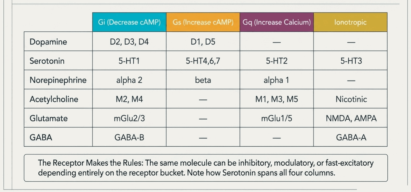
This is the atlas in one grid: every major transmitter system across the top-level receptor buckets. Read across a row to see how one transmitter can pull a neuron in opposite directions depending on which receptor it hits — serotonin is inhibitory at 5-HT₁ autoreceptors (Gᵢ), excitatory-modulatory at 5-HT₂ (Gₜ), and fast-excitatory at the 5-HT₃ ion channel. Read down a column to see which receptors share a signaling logic — and therefore a family resemblance in their pharmacology.
6 · Location, Location, Location: The Serotonin Receptor Map
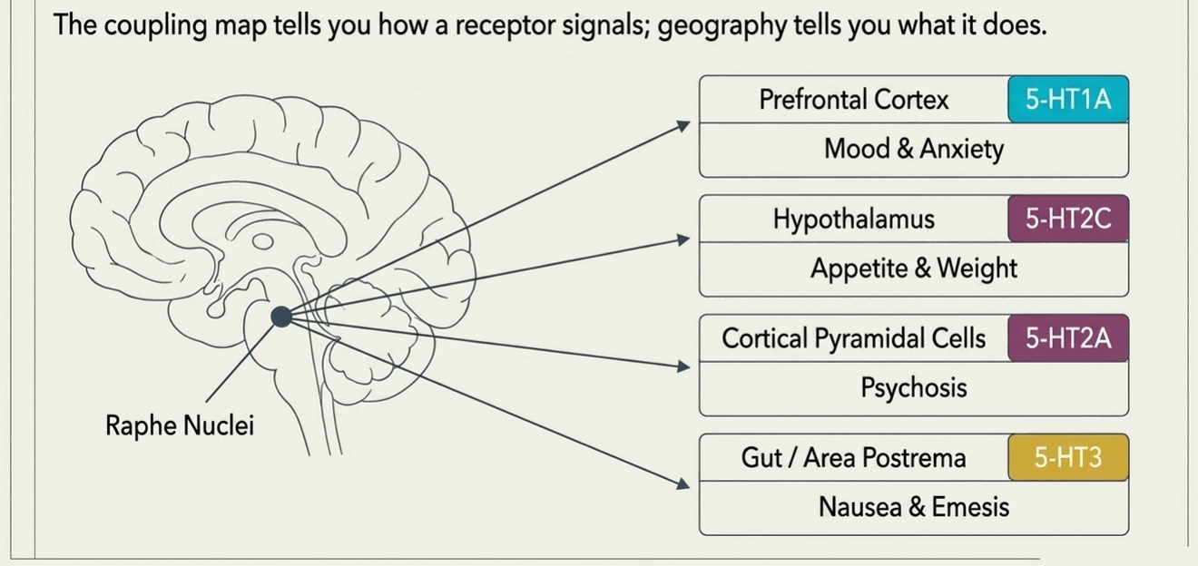
The coupling map tells you how a receptor signals; it does not tell you where it sits — and with serotonin, location is everything. All serotonin originates from a small cluster of raphe nuclei in the brainstem, which project almost everywhere: prefrontal cortex, limbic system, hypothalamus, basal ganglia, brainstem, and gut. The same transmitter therefore produces wildly different effects depending on which receptor, on which cell, in which region it reaches.
The single most clinically important distinction is autoreceptor versus postsynaptic. Serotonin neurons carry inhibitory 5-HT₁₀ autoreceptors on their cell bodies and dendrites (somatodendritic) that slow the neuron’s firing, and 5-HT₁ⁱ/5-HT₁₌ autoreceptors on their terminals that curb release. When an SSRI first blocks reuptake, the extra serotonin lands preferentially on these autoreceptors and actually dampens 5-HT output — one reason antidepressant benefit lags for weeks until those autoreceptors desensitize. Only then does the postsynaptic signal rise. The diagram below maps each receptor to its home.
7 · Muscarinic Receptors, Reconsidered: The Xanomeline Story
For decades the muscarinic system was treated mostly as a liability in psychiatry — the source of the anticholinergic burden (dry mouth, constipation, blurred vision, urinary retention, confusion, and frank delirium) that we try to minimize. The approval of xanomeline–trospium (Cobenfy) in September 2024 flipped that framing: muscarinic agonism is now a validated, first-in-class antipsychotic mechanism — and the first that works without blocking dopamine D₂ receptors at all.
The science is not new; its application is. In the 1990s, Eli Lilly developed xanomeline, an M₁/M₄-preferring agonist, for Alzheimer’s disease. A landmark trial not only slowed cognitive decline but unexpectedly and dramatically reduced psychosis and agitation — the first strong hint that muscarinic activation could be antipsychotic. The program was shelved because peripheral cholinergic side effects were intolerable. Karuna Therapeutics (later acquired by Bristol Myers Squibb) solved that problem elegantly by co-formulating xanomeline with trospium, a peripheral muscarinic antagonist that does not cross the blood–brain barrier — blunting the gut, bladder, and cardiac effects while leaving the central agonism intact.
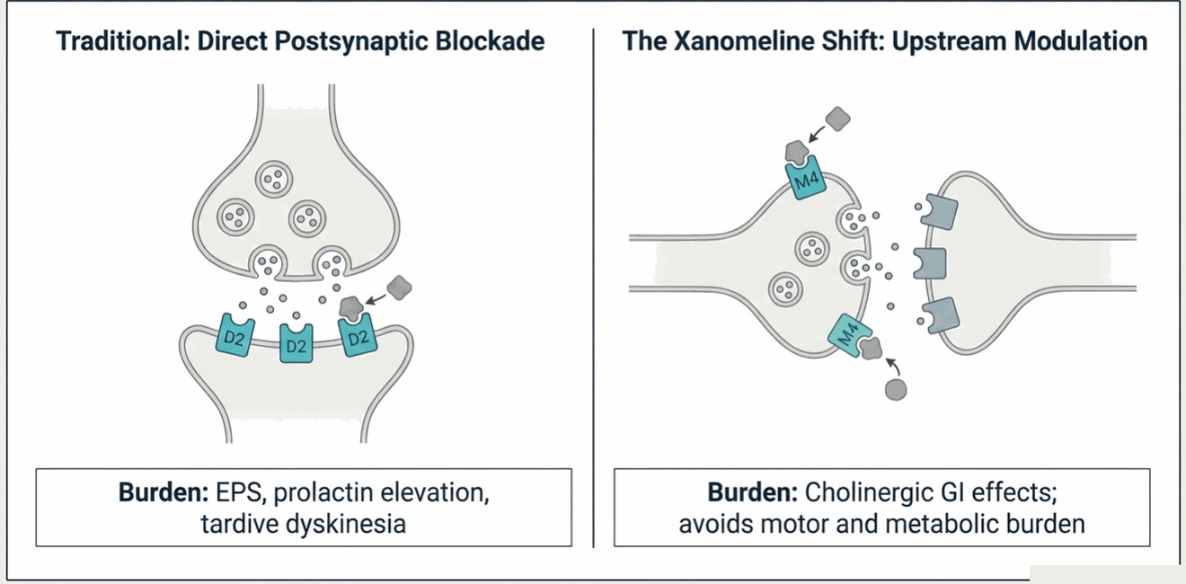
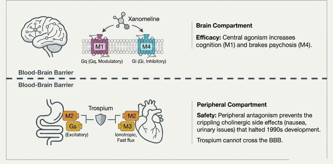
Mechanistically, the two central targets do different jobs. M₄ is enriched in the striatum, where it acts as a brake on dopamine release; stimulating it dampens the excess mesolimbic dopamine signaling thought to drive positive symptoms — but from upstream, rather than by occupying the postsynaptic D₂ receptor. That is why the drug avoids the classic D₂-blockade burden: extrapyramidal symptoms, tardive dyskinesia, and prolactin elevation. M₁, concentrated in cortex and hippocampus, contributes pro-cognitive and antipsychotic effects. In the phase III EMERGENT-2 and EMERGENT-3 trials, xanomeline–trospium separated from placebo on the PANSS by roughly 8–10 points, with a side-effect profile dominated by transient cholinergic GI symptoms rather than metabolic or motor effects.
8 · One Transporter, Region-Dependent Effects
Two facts make norepinephrine pharmacology richer than it looks: transporters are not perfectly selective, and the monoamine systems talk to each other. Together they explain why blocking a single transporter can raise three transmitters, and why the same drug helps different symptoms in different brain regions.
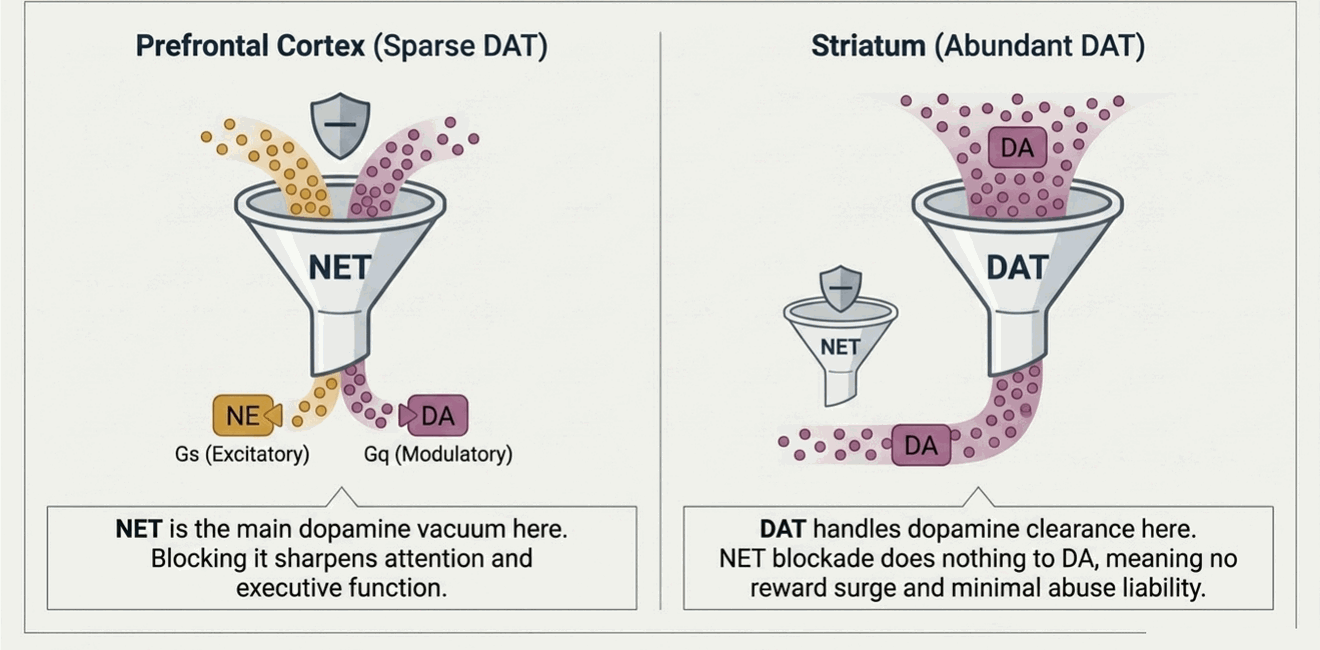
NE reuptake blockade raises dopamine — but only in the cortex
The norepinephrine transporter (NET) also carries dopamine. In most of the brain that is irrelevant, because the dopamine transporter (DAT) does the clearing. But the prefrontal cortex is peculiar: it has very sparse DAT, so cortical dopamine is cleared largely by NET. Block NET and you raise both NE and dopamine in the prefrontal cortex at once. In the striatum and nucleus accumbens — where DAT is dense — NET blockade barely touches dopamine.* That single dissociation explains why atomoxetine (a pure NET inhibitor) sharpens attention and executive function like a stimulant would in the cortex, yet has little abuse liability: it cannot raise accumbens dopamine, so there is no reward surge.
NE reuptake blockade raises serotonin through adrenergic crosstalk
Norepinephrine also steers the serotonin system through two adrenoceptor connections.** Excitatory α₁ adrenoceptors on raphe serotonergic cell bodies mean that more NE tone drives more 5-HT neuron firing (“NE turns on 5-HT release”). Inhibitory α₂ heteroreceptors on serotonin terminals do the opposite — they brake 5-HT release. Mirtazapine is the clean demonstration: it blocks α₂ autoreceptors on NE neurons and α₂ heteroreceptors on 5-HT terminals, disinhibiting both systems without inhibiting reuptake at all.
9 · Go Deeper: The System Deep Dives
Each transmitter system has its own anatomy, receptor nuances, and clinical story. The dedicated chapters below build on this atlas — pathways, receptor-by-receptor pharmacology, and the disorders and drugs tied to each. (Deep-dive chapters are being published in sequence; links activate as each goes live.)
This atlas maps receptors to systems. To see which cell each receptor actually sits on — the neurons and glia, their normal physiology, and how fever, pH shifts, and electrolyte disturbances change their behavior — open the interactive Cellular Neuroscience reference.
Key Takeaways
Three ideas carry most of the weight. First, drugs act at four synaptic nodes — enzymes, transporters, autoreceptors, and postsynaptic receptors — so any mechanism you meet can be located on the synapse diagram. Second, receptors reduce to four signaling buckets, and the bucket predicts both effect and side effect. Third, a single transmitter is neither excitatory nor inhibitory in the abstract; the receptor decides. Hold those three, and the rest of psychopharmacology is detail you can look up — starting with the deep dives above.
Notes & Caveats
* Much of the region-specific dopamine data comes from rodent microdialysis — most notably Bymaster and colleagues (2002) showing that atomoxetine raises dopamine and norepinephrine in the prefrontal cortex but not the striatum or nucleus accumbens. Direct human confirmation is more limited, so treat the magnitude as a well-supported model rather than a precise clinical measurement.
** The α₁/α₂ NE–5-HT crosstalk framing is heavily associated with Stephen Stahl’s teaching model. It is clinically robust and useful for reasoning about drugs like mirtazapine, but it simplifies messier underlying circuitry (multiple receptor subtypes, regional heterogeneity, and feedback loops not shown here).
Hover any * in the text to see its caveat inline, or follow it here.